Ultrasound is one of the gold standard methods for evaluating varicose veins. An ultrasound study produces a comprehensive map of the large and small veins, branching veins, and interconnecting veins of your legs. Ultrasound is often performed as a first step in evaluating varicose veins and also provides valuable follow-up information to monitor the healing process after vein treatment[1]. It can show how well the valves are working and reveal if there is reflux (backup blood flow) or inflammation in a vein[1]. Ultrasound can also show if a vein is partially or completely closed off or if a blood clot has formed.

How Does Ultrasound Work?
Ultrasound is a safe, painless, non-invasive procedure in which high-frequency sound waves are directed toward the veins. As the sound waves meet the structures of the leg, they change frequency depending on the density of each tissue they encounter. The ultrasound machine detects the changing sound wave frequencies and transforms them into a visual image of the underlying structures.
How Does My Doctor Know If I Need an Ultrasound?
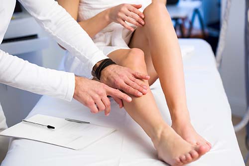
If you have signs or symptoms of venous insufficiency, an ultrasound will likely be required. These include visible varicose or spider veins; swelling of the legs or ankles; itching or aching sensations in the legs; or changes in skin color, particularly development of dark red or bruised appearance in the skin of the ankles and lower legs.
What Can I Expect at My Leg Ultrasound?
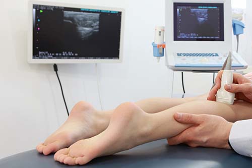
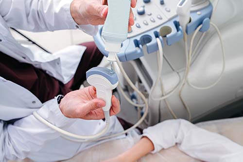
An ultrasound of the legs takes about 30 to 45 minutes. During the procedure, a water-soluble gel is placed on your skin over the area being studied. The ultrasound technician then passes a handheld device across your leg. The device emits ultrasound waves, and the gel helps direct the waves to the veins.
You will likely be placed in both standing and lying down positions during the ultrasound procedure. This ensures the highest quality image in three important ways:
- Helps the ultrasound machine access all the veins.
- Assesses changes in blood flow that might occur in different positions.
- Tests the efficiency of the calf muscle pump, an essential mechanism that assists the flow of blood in the veins through pressure applied by the contraction of the calf muscles.
The ultrasound technician will also apply compression to your thigh and leg, either manually or with a blood pressure cuff, to assess changes in blood flow. Additionally, if a blood clot is suspected in a vein close to the surface or if you are at increased risk for blood clots the technician might gently press on the veins to feel for the presence of a clot.
Typically, the most superficial veins – those closest to the skin surface – are evaluated first, followed by the intermediate veins, and then the deep veins. If you are experiencing swelling or skin changes, such as discoloration, venous eczema, or venous ulcers, examining the deep veins is particularly important as this can reveal if there is compression of a deep vein and it can provide evidence of a previous blood clot [2,3].

How Should I Prepare for My Leg Ultrasound?
Here are a few simple steps you can take before your ultrasound appointment that will help ensure the most accurate ultrasound evaluation of your leg veins:
- Drink plenty of water before your appointment. That way your body will be fully hydrated and the ultrasound will show the peak rates and patterns of blood flow.
- Wear loose-fitting clothing that doesn’t bind or restrict blood flow and that you can remove easily, as needed, for the procedure.
- Avoid using skin lotion on your legs as it can interfere with the ultrasound gel.
How Will My Ultrasound Results Determine What Type of Treatment I Need?
The ultrasound evaluation shows precisely what and where the underlying problem veins are and help your doctor in deciding the best course of therapy for you. For example, it will reveal if you have venous insufficiency due to damaged valves, if there is a blood clot, if one or multiple veins are involved, and where the problem is located. Your recommended treatment will be determined by the cause of the symptoms and the location and extent of the affected veins.
Depending on the ultrasound results your physician may suggest one or a combination of these minimally invasive vein treatments.
VenaSeal™ – Permanently closes off varicose veins with an injection of a medical grade adhesive. If you are experiencing typical varicose vein symptoms such as swelling, itching, numbness, or muscle fatigue VenaSeal™ is a highly effective option. This therapy is also an excellent choice for more advanced vein disease symptoms, including venous ulcers or if your ultrasound reveals vein wall inflammation (thrombophlebitis).
ClosureFast™ – Heat-seals varicose veins with radiofrequency (microwaves). If your ultrasound study shows the involvement of the larger veins ClosureFast™ may be the treatment of choice. This technique has successfully eliminated the long recovery, pain, and bruising associated with conventional vein stripping surgery.
Sclerotherapy – Uses a liquid or foam compound injected into varicose veins that causes them to scar and close. If your vein problems are limited to smaller varicose and spider veins sclerotherapy may be the recommended treatment for you. Results generally take a few weeks to be fully evident as the vein tissue is reabsorbed and new healthy blood flow is established. This technique is often done as a series of treatments.

What Type of Training is Required to Be an Ultrasound Technician?
At Desert Vein and Vascular Institute (DVVI), we take pride in setting and maintaining the highest standards in the industry for ultrasound quality. Obtaining an accurate leg ultrasound is essential to make a precise diagnosis, which helps us determine the most appropriate treatment for each individual. Because we rely on the quality of our ultrasound studies, all our ultrasound technicians are Registered Vascular Technologists (RVT’s) and undergo extensive training on state-of-the-art equipment. The RVT certification raises the standard of vascular ultrasound practice worldwide and promotes best practices for enhanced patient safety. When you come to DVVI for a leg ultrasound you can feel confident that you will receive the highest quality service and the clearest possible images of your leg veins.
Our pursuit of excellence in ultrasound imaging has earned DVVI the distinction of being the only vascular lab in the area to be accredited by the Intersocietal Accreditation Commission (IAC). It is also the reason we were selected as a venous sonography training site by the West Coast Ultrasound Institute’s School of Medical Sonography.
To learn more about how diagnostic ultrasound can assist in your journey to healthy, pain-free legs we invite you to schedule a free consultation today by calling 1-800-827-4267.
Desert Vein and Vascular Institute is the top provider of VenaSeal™, the leading outpatient varicose vein treatment, in the USA. All our physicians are board-certified vascular surgeons who specialize in helping people improve their vascular health.
References
- Doppler ultrasound exam of an arm or leg.
https://medlineplus.gov/ency/article/003775.htm - Diagnosis of venous disease with duplex ultrasound. Phlebology, 2013. 28 Suppl 1
https://www.ncbi.nlm.nih.gov/pubmed/23482553 - Duplex ultrasound evaluation of patients with chronic venous disease of the lower extremities. AJR. American Journal of Roentgenology, 2014. 202(3)
https://www.ncbi.nlm.nih.gov/pubmed/24555602
